

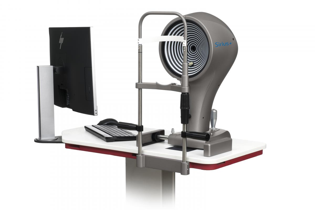
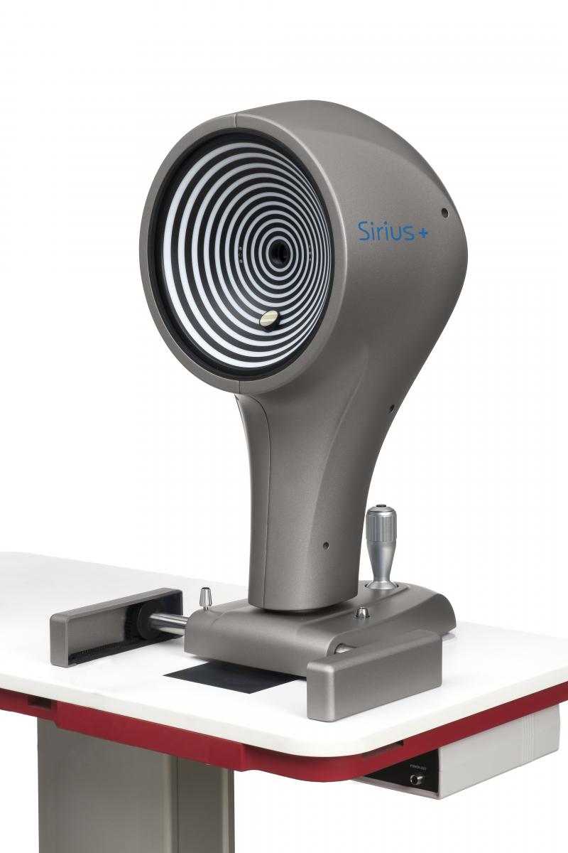
SIRIUS+
Tomograph and corneal topographer
Sirius+ combines Placido Disk topography with Scheimpflug tomography of the anterior segment providing information on pachymetry, elevation, curvature and dioptric power of both corneal surfaces over a diameter of 12 mm. All biometric measurements of the anterior chamber are calculated using up to 100 HR corneal sections. Measurement speed reduces the effect of eye movement producing a high quality accurate measurement.
In addition to the clinical diagnosis of the anterior segment the most common uses are: refractive and cataract surgery, an IOL calculation module is available.
Objective examinations provide an accurate measurment of pupil diameter in scotopic, mesopic and photopic conditions.
KERATOCONOUS SCREENING
Keratoconous screening provides the clinician with important information about the patinets cornea. Understanding this can help prevent complications associated with ectasia before corneal surgery is undertaken.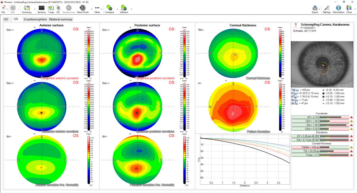
GLAUCOMA SCREENING
For glaucoma specialists Sirius+ enables the measurement of irido-corneal angles and pachymetry. These two values are useful in the diagnosis of the disease.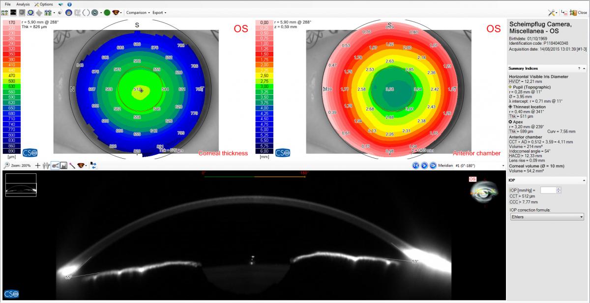
PUPILLOGRAPHY
Sirius+ has built-in pupillography measurement software. The measurement of the pupil in scotopic, mesopic, photopic, conditions and in dynamic mode. knowledge of the center and the diameter of the pupil, is essential for many clinical procedures which seek to optimize vision quality.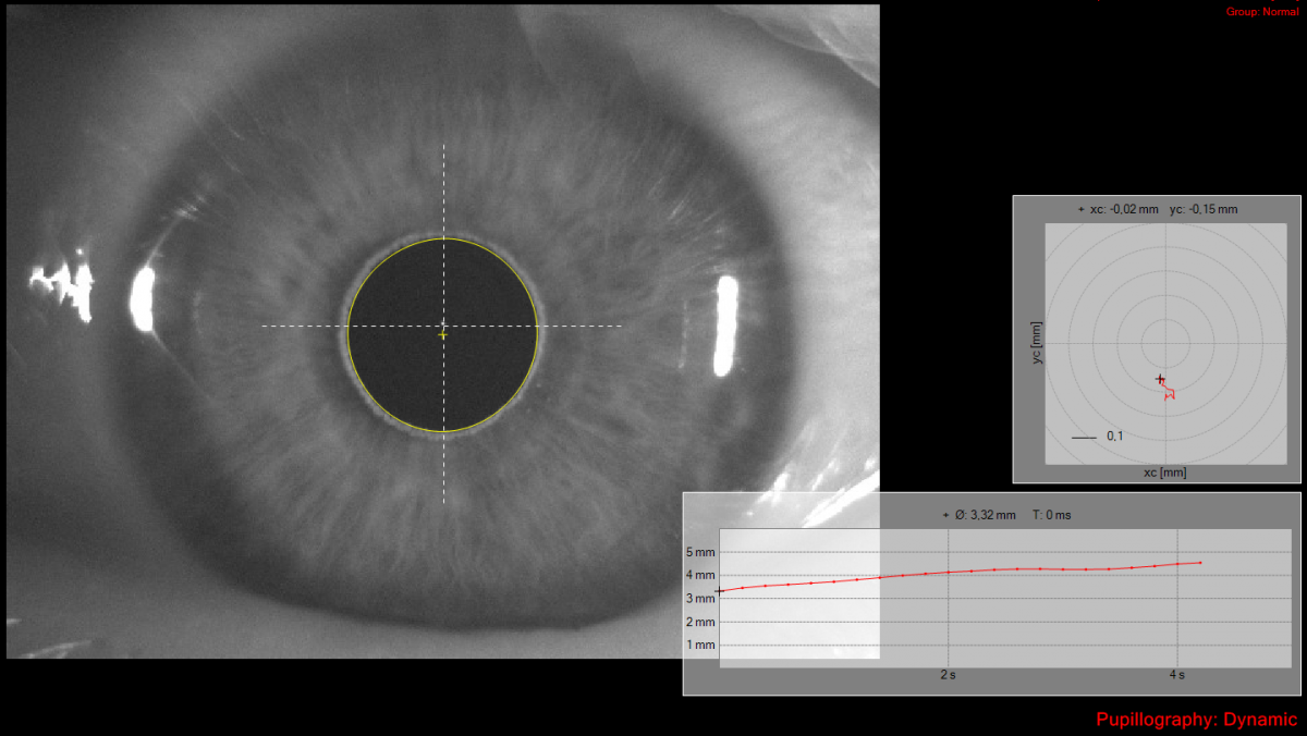
IOL CALCULATION MODULE (optional)
This module is based on Ray-Tracing techniques, regardless of the state of the cornea (untreated or previously treated for refractive purposes), provides the calculation of the spherical and toric power of the intraocular lens.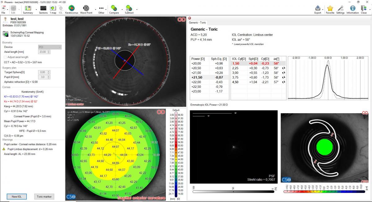
INTRASTROMAL RINGS
On the basis of the pachymetry map and corneal alti- metric data, Sirius+ allows for intrastromal rings system planning, which may be an option for the correction of refractive defects and some forms of keratoconus.
CORNEAL ABERROMETRY
Aberrometric analysis offers a complete overview of the corneal aberrations. It is possible to select the contribution of the anterior, posterior or total cornea for different pupil diameters. The OPD/WFE maps and the visual simulations (PSF, MTF, image convolution with optotype) can help the clinician in understanding or explaining the patient’s visual problems.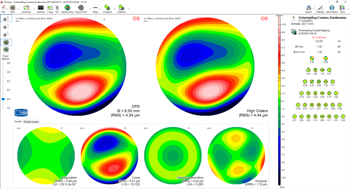
TECHNICAL DATA
| Dimensions (HxDxW) | 509 x 315 x 260mm | |
| Weight | 7Kg | |
| Chin rest movement | 70mm ± 1mm | |
| Minimum height of the chin cup from table | 24cm | |
| Base movement (xyz) | 105 x 110 x 30mm | |
| Working distance | 74mm | |
| LIGHT SOURCES | ||
| Placido disk | LED @400-700nm | |
| Scheimpflug | LED @475nm UV-free | |
| Pupillography | LED @940nm | |
| Fluoresceine lighting | LED @470nm | |
| Auxiliary lighting | LED 400-700nm | |
| TOPOGRAPHY | ||
| Placido disk rings | 22 | |
| Measured points | from 42032 to 151232 for the front surface from 36400 to 145600 for the rear surface | |
| Topographic covering | 12mm | |
| Dioptric measurement range | 1D to 100D | |
| Measurement accuracy | Class A according to the UNI EN ISO 19980-2012 | |
| Compatibility with standard | DICOM v3 (IHE integration profile EYECARE Workflow) | |
| ACCESSORIES | ||
| Light diffuser filter for auxiliary illumination, magnetic lock | light diffuser filter | |
| Yellow barrier filter, magnetic lock | 530 nm filter | |
| Additional lens, magnetic lock | -6D lens | |
| Calibration tool | r 8 mm calibration tool | |
| MINIMUM SYSTEM REQUIREMENT | ||
|
PC: CPU: I3 or higher ( suggested I5) - CHIP SET: intel - RAM: 4 Gbyte or higher (suggested 8 Gbyte) -
|
||